Synapse imaging reveals a big role for small protrusions Identification of a new mechanism governing synapse formation

Neurons in the brain are wired to each other through the connections called “synapses”. During the first few years of infancy, humans acquire myriad of skills to walk, to communicate and to socialize. Such highly sophisticated skills can be only achieved through precise synapse formation.
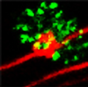
“Small protrusions” from an axon of a cerebellar granule cell at the onset of synapse formation
© Aya Ito-Ishida
The small protrusions changed shape and surrounded the postsynaptic structures of a Purkinje cell, which was associated with the accumulation of postsynaptic glutamate receptor delta 2 (green). The finding suggested that the small protrusions are essential for the functional maturation of the synapses. This microscopic image was obtained from a live cerebellar slice of a Cbln1-lacking mouse, which had been treated with recombinant Cbln1 for seven hours prior to image acquisition. The granule cells and the glutamate receptor delta 2 were visualized by fluorescent proteins of different colors.
A research group including Prof. Shigeo Okabe and Dr. Aya Ito-Ishida of the University of Tokyo Graduate School of Medicine, Department of Cellular Neurobiology and Prof. Michisuke Yuzaki from Keio University have discovered a new mechanism that governs synapse formation. The group has for the first time directly observed the entire process of synapse formation in detail in live neurons in the mouse cerebellum, a part of the brain that controls body movement. It was found that the neurons extend “small protrusions” to promote immature synapses that mature and become functional. These “small protrusions” were formed through the interaction of three proteins, Cbln1, glutamate receptor delta 2, and neurexin, all of which are essential for the formation of synapses in the cerebellum.
Recent advances in research have revealed that several types of developmental neurological disorders including autism are due to the failure of normal synapse development. The discovery of these “small protrusions” may open new avenues to the basic research in synapse development and neurodevelopmental disorders.
Press release [PDF] (Japanese)
Paper
Aya Ito-Ishida, Taisuke Miyazaki, Eriko Miura, Keiko Matsuda, Masahiko Watanabe, Michisuke Yuzaki, Shigeo Okabe,
“Presynaptically released Cbln1 induces dynamic axonal structural changes by interacting with GluD2 during cerebellar synapse formation,
Neuron 2012/11/8, doi: 10.1016/j.neuron.2012.07.027.
Article link
Links
Graduate School of Medicine and Faculty of Medicine
Department of Cellular Neurobiology
Keio University, School of Medicine, Department of Physiology


