Temporal jitter of observed signal drastically improves fMRI resolution Simultaneous observation of neural and vascular activity

Many recent discoveries on the human brain are based on observation of neural activity with functional magnetic resonance imaging (fMRI). fMRI provides a non-invasive mean of measuring changes in oxygen concentrations in the brain, and is widely used to estimate brain activity under the assumption that neural activity gives rise to a characteristic temporal response in the blood-oxygen-level-dependent (BOLD) signal. However, while neural activity takes place on a millisecond timescale, current fMRI technology can only offer temporal resolution of multiple seconds. In addition, the spatial resolution of fMRI is on the millimeter scale. Improvement in both spatial and temporal resolution is required for further understanding of the human brain.

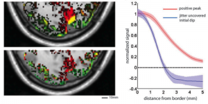
© Masataka Watanabe, fMRI brain activity evoked by two adjacent visual stimuli (green/red). Upper-left: analysis using positive BOLD. Lower-left: analysis using jitter-uncovered initial dip. Right: spatial spread of the two signals.
The current limit of fMRI spatiotemporal resolution is related to the type of signals that are observed. There are two signals related to increased neural activity in the brain: an initial dip in oxygen concentration, and a later positive peak in oxygen concentration. The sharp initial dip reflects increase of oxygen consumption as a result of neural activity, while the later positive peak signal captures the vascular response. the majority of current fMRI studies use the later positive peak due to the difficulty in capturing the initial dip. Robust observation of the initial dip would enable a great increase in fMRI spatiotemporal resolution.
A research team lead by Professor Masataka Watanabe at the University of Tokyo Graduate School of Engineering’s Department of Systems Innovation has shown that the positive peak of the stimulus evoked BOLD signal exhibits a trial-to-trial temporal jitter in the order of seconds. Moreover, the researchers showed that this trial-to-trial variability was the main cause for the difficulty in observing the initial dip and can be exploited to uncover it. Initial dips were observed in the majority of voxels by pooling trial responses with large peak latencies. Initial dips exposed by this procedure possess higher spatial resolution compared to the positive BOLD signal in the human visual cortex. These findings allow for the reliable observation of fMRI signals that are physiologically closer to neural activity, leading to improvements in both temporal and spatial resolution. In principle, it permits measurements with a spatial resolution of 100 micrometers and temporal resolution of 100 milliseconds. The method should enable the construction of a far more detailed map of brain activity and fine structure of brain function, leading to the elucidation of higher brain functions such as language, logic and intuition. Together the method can be applied to medical diagnosis for simultaneous observation of neural and vascular state.
This research was published online in Current Biology on October 17, 2013 at 12:00 (EST).
Paper
Masataka Watanabe, Andreas Bartels, Jakob Macke, Yusuke Murayama, Nikos Logothetis,
“Temporal jitter of the BOLD signal reveals a reliable initial dip and improved spatial resolution”,
Current Biology Online Edition: 2013/10/17 (EST), doi: 10.1016/j.cub.2013.08.057.
Article link
Links
Graduate School of Engineering
Department of Systems Innovation, Graduate School of Engineering


