Visualization of the movement of calcium ions in neurons The endoplasmic reticulum as pipeline and store for calcium ions


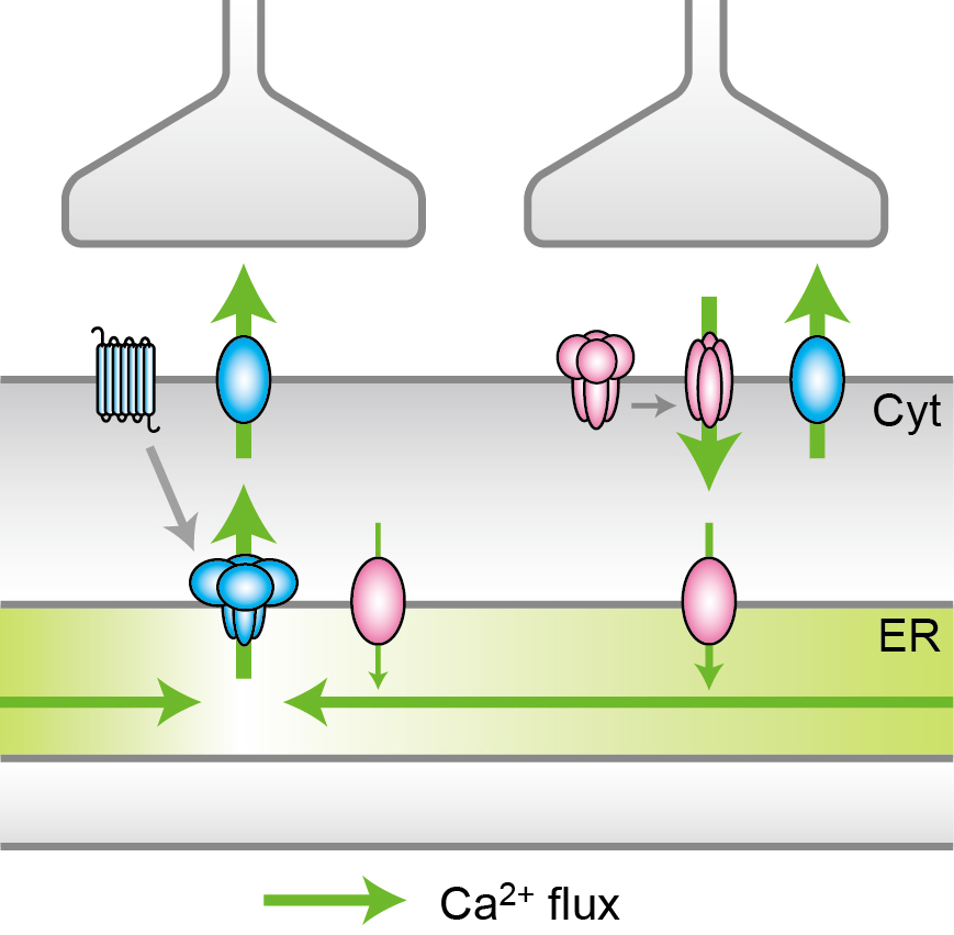
Ca2+ flux in response to two kinds of synaptic inputs
(Left) A synaptic input induces the local depletion of Ca2+, which is followed by diffusion from the surrounding region. (Right) Another synaptic input induces the uptake of Ca2+ from the cytoplasm (Cyt) to ER via Ca2+ pump, followed by the accumulation and the maintenance of Ca2+ within ER.
© 2015 Iino Lab, The University of Tokyo.
University of Tokyo researchers have successfully visualized the movement and accumulation of calcium ions (Ca2+), a major intracellular messenger, within the endoplasmic reticulum (ER), an intracellular organelle, revealing subcellular responses to synaptic inputs in neurons. This visualization technique is expected to be useful for the investigation of disease mechanisms and the development of therapeutic drugs.
Calcium ions are involved in the regulation of various functions including muscle contraction and learning and memory. ER, which appears as a fine tubular network expanded throughout the cell, is a major source of calcium ions. Calcium ions are stored in high concentrations within the ER and is released in response to extracellular stimulation. However, the dynamics of calcium ions within the ER remains poorly understood due to a lack of appropriate techniques for observation and measurement.
In this study, Professor Masamitsu Iino, Lecturer Yohei Okubo and their colleagues at the Graduate School of Medicine’s Department of Pharmacology have developed a novel fluorescence probe G-CEPIA1er to visualize calcium ions within ER in neurons. Analysis of fluorescence images revealed the movement of calcium ions from a replete region to a depleted region upon a synaptic input. Another synaptic input induced the accumulation and the short-term maintenance of calcium ions within ER. These results revealed that neuronal response to synaptic inputs through ER functions as a pipeline and as a volatile memory.
“Revealing the basics of calcium ion regulation mechanisms not only improves our understanding of cellular function but also the possibility for the clarification of disease mechanisms,” says Lecturer Okubo. He continues, “For example, it is suggested that the disruption of intracellular calcium homeostasis including in the ER is involved in neurodegenerative diseases.”
Paper
, "Visualization of Ca2+ filling mechanisms upon synaptic inputs in the endoplasmic reticulum of cerebellar Purkinje cells", The Journal of Neuroscience 35(48) 15837-15846: 2015/12/2 (Japan time), doi: 10.1523/JNEUROSCI.3487-15.2015.
Article link (Publication)
Links
Department of Cellular and Molecular Pharmacology, Graduate School of Medicine


