

Curiosity and Innovation
Not Just GPS:
Basic Physics Research Opening Doors to New Worlds
[High-tech Treatments] Particle Research and Medicine
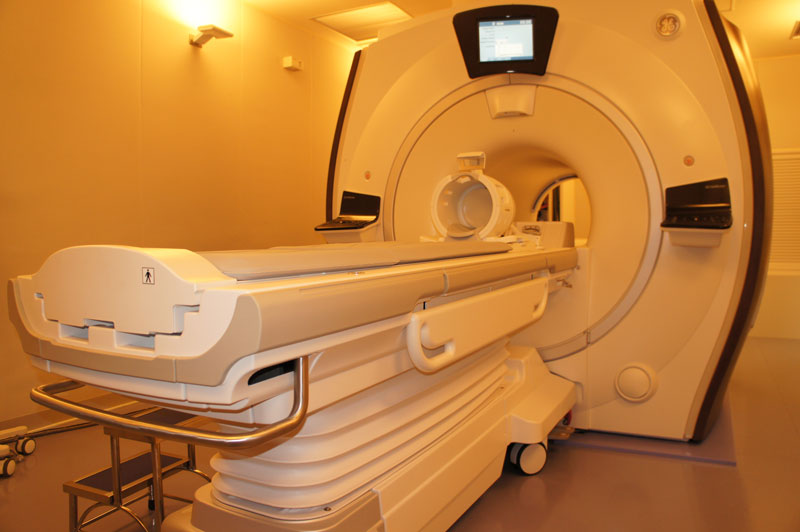
Since they emit strong magnetic fields, MRI scanners are placed in dedicated rooms. It is strictly prohibited to bring in any metallic objects or magnetic cards.
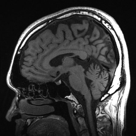
The imaging technology in MRI scans allows them to see through the skull more clearly than X-ray CT scans, which are more obstructed by bone. This ability makes MRI scans ideal for examinations of the brain.
Machines that have been devised and developed based on fundamental particle physics research are essential in the contemporary medical setting. These machines are tomographic imaging devices that would be familiar to anyone who has undergone a medical checkup, such as CT (X-ray computed tomography) and MRI (magnetic resonance imaging) scanners. Although to the lay person these devices may conjure up images of a “super-powered X-ray machine,” what is the reality of such equipment? We put this question to Professor Akira Kunimatsu.
“X-ray computed tomography (CT) is an imaging technique whereby the body is scanned from all angles (360 degrees) with X-rays, which are then computer-processed to see the extent to which the X-rays have been absorbed in each direction. Magnetic resonance imaging (MRI) on the other hand uses a physical phenomenon known as nuclear magnetic resonance (NMR). When radio waves at more or less the same frequency as FM radio are passed through the hydrogen atoms present in the body, the hydrogen nuclei send back a signal. The signal that is sent back differs depending on the type of material that is being scanned, and a computer is used to analyze the signal, as well as its frequency and phase, which makes it possible to create an image of what kinds of materials are in the body and where. For example, in the case of the brain, it is possible to identify grey matter that contains many nerve cells, or white matter that contains many nerve fibers.”
Unlike X-ray CT scans, a major characteristic of MRI is that the subject is not exposed to radiation and it is possible to create an image that details anatomical composition clearly. However, an MRI examination takes longer than a CT scan and typically only larger hospitals can afford to have MRI devices. Therefore, the general rule for most medical conditions is that first an X-ray CT scan is conducted and if this shows some abnormality, a further, more detailed MRI is administered. However, recently medical practice is seeking to minimize the radiation to which patients are exposed, so there are increasing numbers of cases where MRI is used as the first option.
“It was a truly revolutionary development in medicine to be able to look from the outside and see inside the body in detail. The next challenge will be to achieve imaging not only of shapes and forms but also of actual functions. It is difficult to determine whether something in the body has shrunk because organ function has decreased, or whether function has decreased because the organ has shrunk. It would be a great innovation in medicine if we were to be able to achieve better imaging of both form and function in the human body.”

Akira Kunimatsu
Associate Professor of Radiology, Graduate School of Medicine
http://www.ut-radiology.umin.jp/