Simple test can find rare cause of lower back pain Doctors hope to give patients correct diagnosis


About 20 percent of patients with long-term low back pain receive the non-diagnosis of nonspecific or idiopathic pain – aching with no known cause. Researchers hope their simple technique will allow doctors to find a specific diagnosis for some patients.
Medical experts from the University of Tokyo have identified a simple test doctors can use to diagnose a rare condition that causes extreme back pain.
Patients can receive a common imaging test, an MRI (magnetic resonance imaging), twice: once while lying flat on their backs (supine) and once while lying face-down on their stomachs (prone). Doctors then compare images of the two positions to identify unusual movement of the nerves branching off from the spinal column.
"This test is noninvasive and simple to perform. We are confident that our methodology has the potential to become a critical diagnostic tool for some patients searching for the cause of their low back pain," said Dr. Masahiko Sumitani from the University of Tokyo Hospital, an author of the recent research paper in Pain Practice.
Although there are many common reasons for back pain, about 20 percent of patients with long-term low back pain receive a non-diagnosis of nonspecific or idiopathic pain – aching with no known cause. Other times, patients are told their back pain is caused by mental illness.
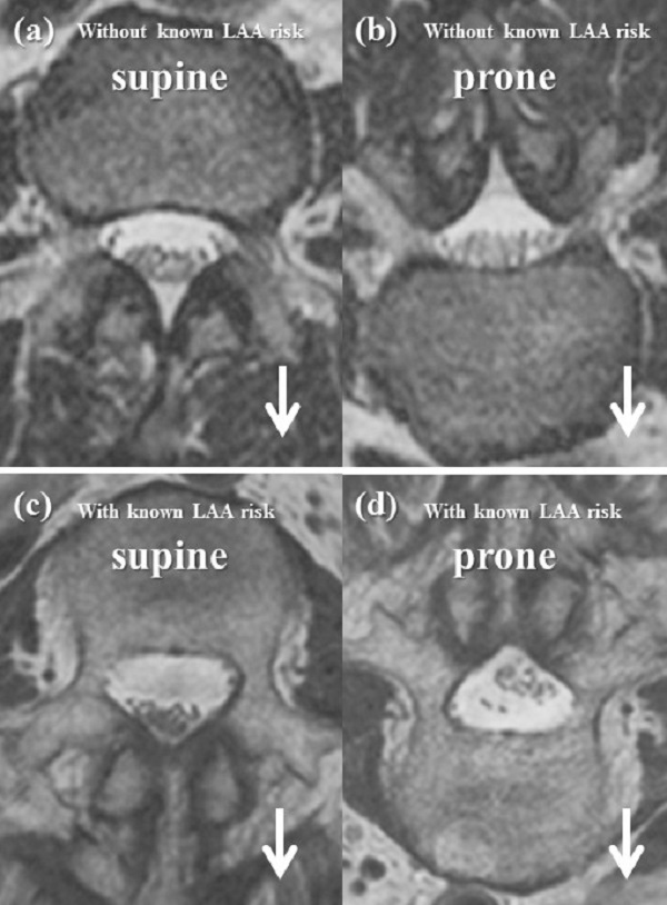
The upper panel (a and b) depicts images from a single patient without known risk factors for lumbar adhesive arachnoiditis. The lower panel (c and d) depicts the results for a single patient with a known risk factor (i.e., lumbar spinal surgery) for the condition. Downward arrows indicate the direction of gravity. © 2019 Masahiko Sumitani. Originally published in Pain Practice.
Normally, a thin, spider-weblike membrane wraps and protects the nerves all along the spine. Sometimes due to injury, chemical exposure, or a complication from prior spinal surgery, that gossamer membrane grows thick and sticks to other structures in the space around the spinal column of the lower back. Then, the nerves that branch off from the spinal column cannot be pulled downward by gravity as they should be during normal physical activity, thereby creating pain.
The condition, lumbar adhesive arachnoiditis, is named for that spider web membrane (arachnoiditis) sticking (adhesive) to other structures in the lower back portion of the spine (lumbar). Lumbar adhesive arachnoiditis affects 25,000 people worldwide each year, according to Orphanet, a French-led global initiative that collects information on rare diseases.
"Lumbar adhesive arachnoiditis is a debilitating neuropathic condition. However, the medical community has not established criteria to diagnose patients with the condition, and therefore, most physicians are probably still unaware of this disease," said Sumitani.
The research team's MRI study revealed that doctors can visualize where the nerves are positioned within the membranes of the spinal column and potentially identify patients at risk for lumbar adhesive arachnoiditis.
Researchers hope that their technique will become well known within the medical community and allow some patients to finally receive an accurate diagnosis of the cause of their low back pain.
"For patients with so-called nonspecific low back pain, our findings reveal that a physically 'specific' cause may exist, and thus their treatment may be improved," said Sumitani.
Collaborators from the Imaging Center at Ochanomizu Surugadai Clinic in Tokyo contributed to this research.
Papers
Rikuhei Tsuchida, Masahiko Sumitani, Kenji Azuma, Hiroaki Abe, Jun Hozumi, Reo Inoue, Yasushi Oshima, Shuichi Katano, Yoshitsugu Yamada, "A Novel Technique Using Magnetic Resonance Imaging in the Supine and Prone Positions for Diagnosing Lumbar Adhesive Arachnoiditis: A Preliminary Study," Pain Practice: July 20, 2019, doi:10.1111/papr.12822.
Link (Publication )
)


