


PRESS RELEASES

Memory, learning and decision-making studied in worms Researchers identify role of interneurons in food-seeking behavior Research news
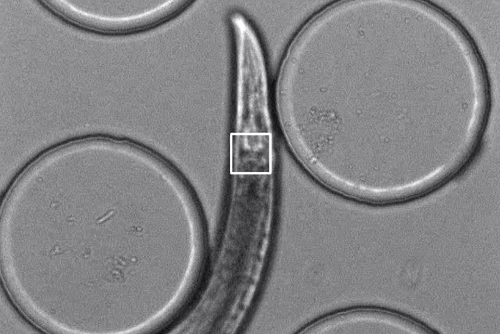
Three views of the same recording show a worm starting in an area of higher salt concentration, moving towards a lower salt concentration, stopping, and then changing directions to go back towards the higher-salt area. The dark circles are pillars in the container that act like speed bumps, slowing down the worm so the microscope can track its movements. Note how the GcaMP6s signal (middle) becomes brighter when the worm reverses direction. Left: C. elegans viewed under normal white light with a square drawn around the neurons examined in the research. Middle: Fluorescent light view of genetically encoded GCaMP6s, an indicator that a neuron is sending a signal to another neuron. Right: Fluorescent light view of mCherry, a fluorescent tag added to the same neuron as GcaMP6s. © Video by Project Researcher Hirofumi Sato, CC BY 4.0
As anyone who has ever procrastinated knows, remembering that you need to do something and acting on that knowledge are two different things. To understand how learning changes nerve cells and leads to different behaviors, researchers studied the much simpler nervous system of worms.
“In this study, we can now translate neuronal activity to behavioral response,” said Project Researcher Hirofumi Sato, a neuroscientist at the University of Tokyo and first author of the research paper recently published in Cell Reports.
The discovery was made possible using technology that researchers describe as a “robot microscope,” first developed in 2019 by researchers at Tohoku University in Miyagi Prefecture, northeastern Japan.
The technique involves genetically modifying the worms to add fluorescent tags onto specific molecules. The microscope then detects and tracks the fluorescent light as a worm crawls around, meaning researchers can watch chemical signals travel through and between individual neurons in awake, unrestrained animals.
The worms used in research studies, C. elegans, don’t eat pure salt, but researchers can train worms to associate high or low salt levels in their environment with food. When transferred to any new environment, trained worms will begin searching for food using salt levels as a clue about which direction they should go. For example, if worms learned to expect food in high-salt areas but they notice that salt levels are decreasing as they travel, the worms will stop and change directions to try to find a higher salt level. With additional training, worms can also learn the opposite food-salt level association.
Neuroplasticity, or the brain’s ability to change and “rewire” neurons, is essential for any learned behavior. The mystery for the scientific community is how different environmental clues (high or low salt) can lead to the same physical behavior (stop and change direction).
“Many animals show this flexible learned behavior pattern, so we want to understand the mechanism,” said Sato.
This type of behavior requires a sensory neuron (which detects salt), motor neurons (which control movement) and interneurons (which communicate between the other two types). Although C. elegans only have 302 neurons in their entire 1-centimeter-long bodies, these same types of neurons exist in humans and communicate using the same signal molecule.
Specifically, that signal molecule is glutamate, widely recognized as one of the brain’s most important signaling molecules.
“We know that if there is a defect in glutamate signaling, that might cause Alzheimer’s disease or other neuronal diseases,” said Sato.
The UTokyo team’s new data found two different types of glutamate receptors on the same interneuron are involved in the worms’ behavior. Both inhibitory and excitatory glutamate receptors respond in the same pattern, but at different intensities based on whether the worms had learned to seek high or low salt concentrations.
The exact mechanism controlling the motor neuron’s signals to the interneuron’s glutamate receptors remains unclear. However, this is one of the first documentations of glutamate signaling between sensory and interneurons showing experience-dependent plasticity.
Future research will continue to investigate exactly how the sensory neuron and interneuron communicate.
Papers
Hirofumi Sato, Hirofumi Kunitomo, Xianfeng Fei, Koichi Hashimoto, and Yuichi Iino, "Glutamate signaling from a single sensory neuron mediates experience-dependent bidirectional behavior in Caenorhabditis elegans," Cell Reports: May 25, 2021, doi:10.1016/j.celrep.2021.109177.
Link (Publication )
)


