


PRESS RELEASES

New imaging technique is no last resort Researchers created a new and improved way to view the mechanics of life Research news
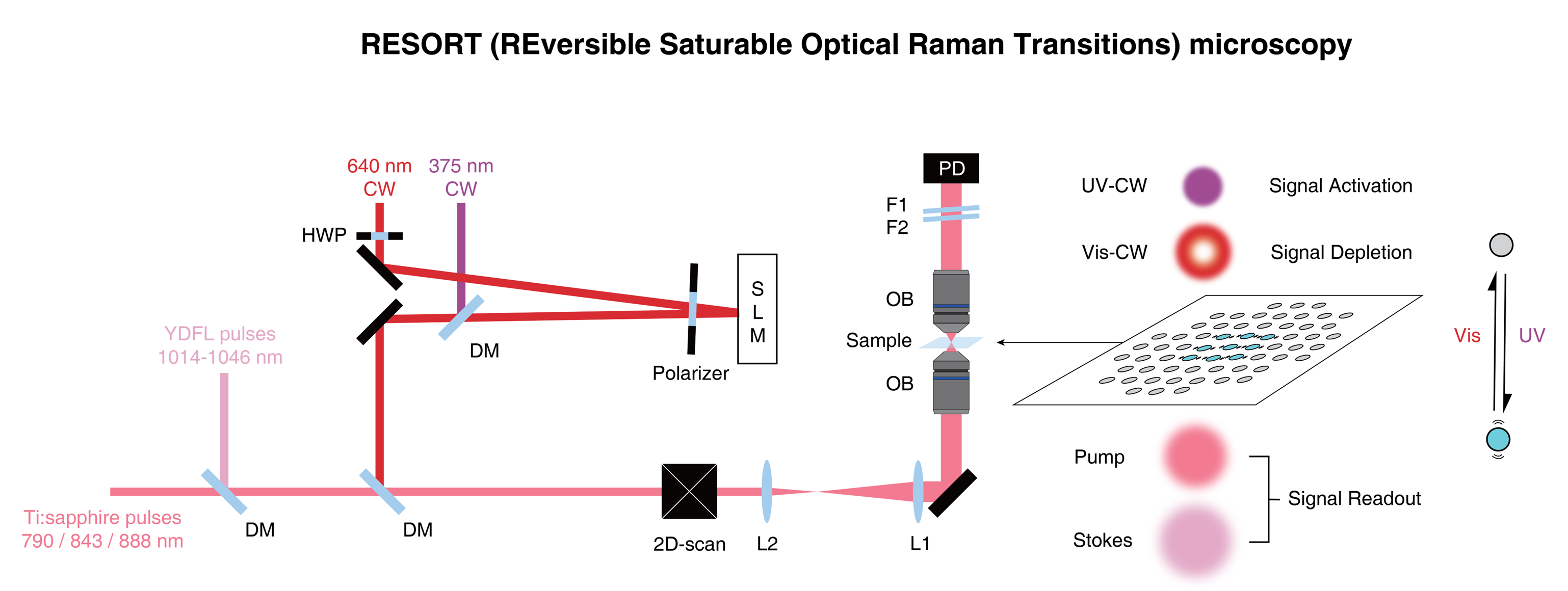
RESORT. A diagram to show the basic overview of the system. Firstly, the sample is labeled with the photoswitchable Raman probe. It’s then irradiated with two-color infrared laser pulses, ultraviolet light, and a special donut-shaped beam of visible light to constrain the area where Raman scattering can occur. As a result, the probe can be detected at a very precise point for high spatial resolution images. ©2023 Ozeki et al. CC-BY
There are various ways to image biological samples on a microscopic level, and each has its own pros and cons. For the first time, a team of researchers, including those from the University of Tokyo, has combined aspects from two of the leading imaging techniques to craft a new method of imaging and analyzing biological samples. Its concept, known as RESORT, paves the way to observe living systems in unprecedented detail.
For as long as humanity has been able to manipulate glass, we have used optical devices to peer at the microscopic world in ever increasing detail. The more we can see, the more we can understand, hence the pressure to improve upon tools we use to explore the world around, and inside, us. Contemporary microscopic imaging techniques go far beyond what traditional microscopes can offer. Two leading technologies are super-resolution fluorescence imaging, which offers good spatial resolution, and vibrational imaging, which compromises spatial resolution but can use a broad range of colors to help label many kinds of constituents in cells.
“We were motivated by the limitations of these kinds of imaging techniques to try and create something better, and with RESORT we are confident that we have achieved this,” said Professor Yasuyuki Ozeki from the University of Tokyo’s Research Center for Advanced Science and Technology. “RESORT stands for reversible saturable optical Raman transitions, and it combines the benefits of super-resolution fluorescence and vibrational imaging without inheriting the detriments of either. It is a laser-based technique that uses something known as Raman scattering, a special interaction between molecules and light which helps identify what’s in a sample under the microscope. We successfully performed RESORT imaging of mitochondria in cells to validate the technique.”
There are several stages to RESORT imaging, and although it might seem complicated, the setup is less complicated than that of the techniques it’s aiming to replace. Firstly, the specific components of the sample to be imaged need to be labeled, or stained, with special chemicals called photoswitchable Raman probes, whose Raman scattering can be controlled by the different kinds of laser light employed by RESORT. Next, the sample is placed within an optical apparatus used to correctly illuminate the sample and build an image of it. For that to occur, the sample is then irradiated with two-color infrared laser pulses for detecting Raman scattering, ultraviolet light and a special donut-shaped beam of visible light. Together, these constrain the area where Raman scattering can occur, which means the final stage, imaging, can detect the probe at the very precise point, which leads to a high spatial resolution.
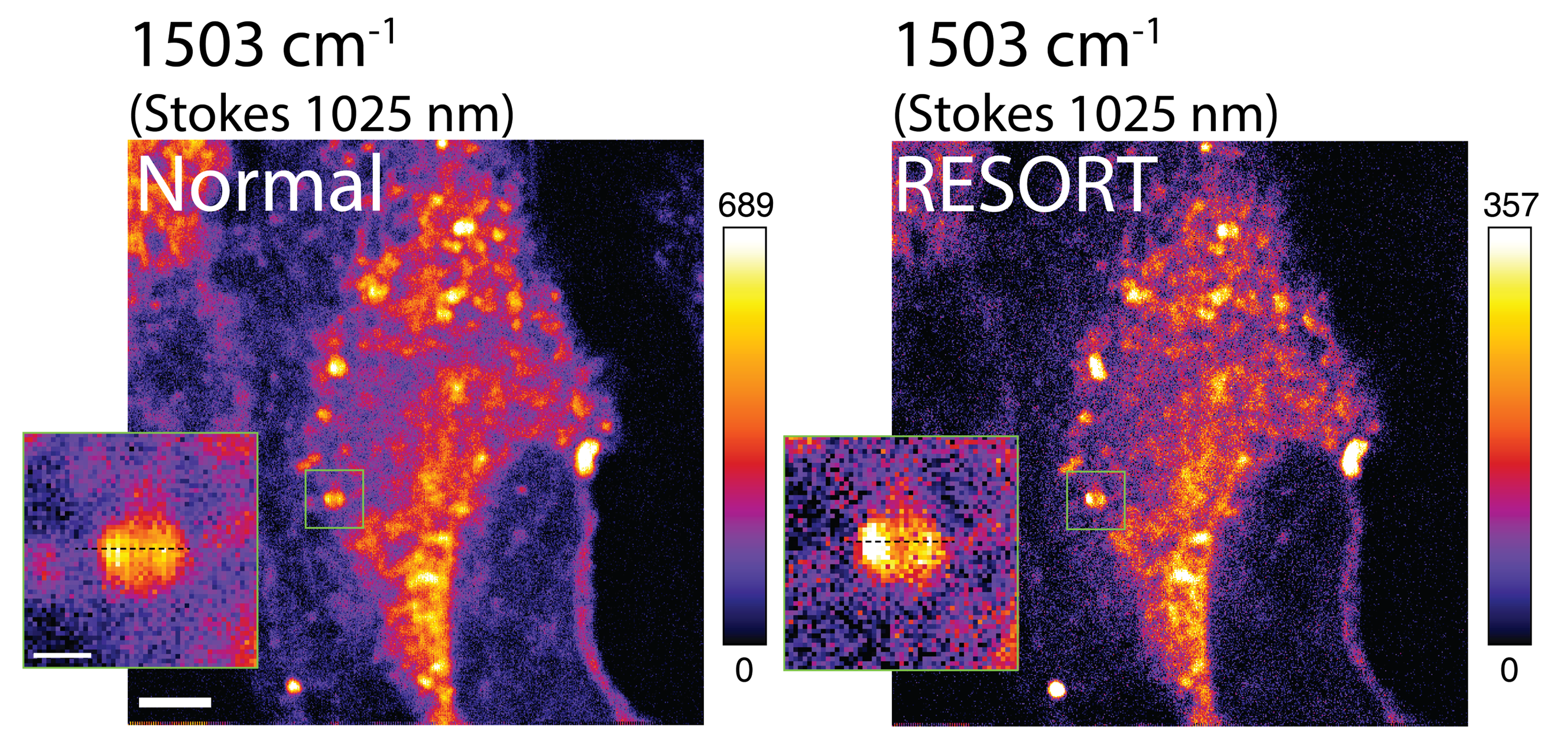
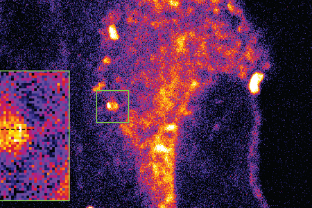
Living cells under the microscope. A comparison of RESORT and a prior imaging technique known as stimulated Raman scattering (SRS). While the difference may look subtle at first glance, the sharper image offered by RESORT can be of great benefit to researchers identifying and labeling components of cells, such as mitochondria. ©2023 Ozeki et al. CC-BY
“It’s not just about gaining higher-resolution images of microscopic samples; after all, electron microscopes can image these things in far greater detail,” said Ozeki. “However, electron microscopes necessarily damage or impede the samples they observe. Through the future development adding more colors to the palette of Raman probes, RESORT will be able to image many components in living samples in action to analyze complex interactions like never before. This will contribute to a deeper understanding of fundamental biological processes, disease mechanisms and potential therapeutic interventions.”
The team’s main aim was to improve microscopic imaging for use in the medical research field and related areas. But the advancements it has made in the design of the laser could be used in other laser applications as well, where high power or precise control is required, such as materials science.
Papers
Jingwen Shou, Ayumi Komazawa, Yuusaku Wachi, Minoru Kawatani, Hiroyoshi Fujioka, Spencer John Spratt, Takaha Mizuguchi, Kenichi Oguchi, Hikaru Akaboshi, Fumiaki Obata, Ryo Tachibana, Shun Yasunaga, Yoshio Mita, Yoshihiro Misawa, Ryosuke Kojima, Yasuteru Urano, Mako Kamiya & Yasuyuki Ozeki, "Super-resolution vibrational imaging based on photoswitchable Raman probe," Science Advances: June 16, 2023, doi:10.1126/sciadv.ade9118.
Link (Publication )
)
Related links
- Research Center for Advanced Science and Technology
- Department of Electrical Engineering and Information Systems
- Graduate School of Pharmaceutical Sciences


