Gene micro-determination in micro-dissected tissue Genes activated in brain follow a power law

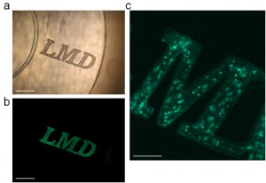
A representative example how elaborately a tissue specimen can be cut out from a fluorescent Nissl-stained biological tissue using a laser beam
© Yoshioka, W. et al. Sci. Rep. 2:783, 2012
We first wrote the letters ‘LMD’ in a tissue specimen having each letter connected to each other. Then, the region of interest composing of a shape of “LMD” was cut-out using the laser beam, collected into the cap of the sampling tube, and visualized under a light microscope (a) and a fluorescence microscope (b, c). Scale bars = 200 μm (a, b) and 50 μm (c).
A histoanatomical context is imperative in an analysis of gene expression in a cell in a tissue to elucidate physiological function of the cell. A research group, composed of Wataru Yoshioka, Masaki Kakeyama, Chiharu Tohyama and other researchers at the Laboratory of Environmental Health Sciences in the Graduate School of Medicine of the University of Tokyo, in collaboration with Ryutaro Nishiyama at Leica Microsystems K. K., made technical advances in laser microdissection (LMD) in combination with the absolute quantification of small amounts of mRNAs from a region of interest (ROI) in fluorescence-labeled tissue sections. This newly-developed fluorescence LMD real-time quantitative polymerase chain reaction (PCR) method has three orders of dynamic range, and is capable of determining absolute amounts of RNAs, with the lower limit of ROI-size corresponding to a single cell.
For its application, the researchers determined the mRNA abundance in the brains of mice. It has been known when mice are transferred from their home cage to a novel environment, neurons are activated in a specific brain area. The brain was collected from the mice transferred to a novel environment and frozen immediately until LMD analysis. The hippocampal regions, CA1, CA2, and DG, were identified by morphological characteristics, and tissue fragments of each region, containing approximately a few hundred neurons, were subjected to real-time quantitative PCR for the determination of analysis of immediate early genes (IEGs), used as surrogate markers for neuronal activity. The hippocampus plays an important role in memory, and is associated with stress responses. In this study, when researchers studied the relationship of the average basal abundance of each IEG with the fold increase in gene expression, i.e., a ratio of the average abundance of the same IEG in the novel environment group over the home cage group, they found that the expression of the IEGs has a distinct activation profile in region CA-1 but not in CA2 or DG. That is, the relationship found in the CA-1 was found to conform a power law distribution.
The use of this method in future studies may lead to the discovery of novel phenomena and deepen our understanding on the physiological significance of gene expression in ROIs in a variety of organs and tissues.
Press release [PDF] (Japanese)
Paper
Wataru Yoshioka, Nozomi Endo, Akie Kurashige, Asahi Haijima, Toshihiro Endo, Toshiyuki Shibata, Ryutaro Nishiyama, Masaki Kakeyama, Chiharu
Tohyama,
“Fluorescence laser microdissection reveals a distinct pattern of gene activation in the mouse hippocampal region”,
Scientific Reports Online Edition: 2012/10/30 PM7:00(Japan time), doi: 10.1038/srep00783.
Article link
Links
Center for Disease Biology and Integrative Medicine
Laboratory of Environmental Health Sciences (Japanese)


