


A new image of hearing Researchers can now see every cell for hearing in 3D
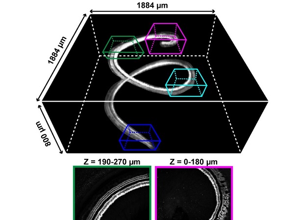
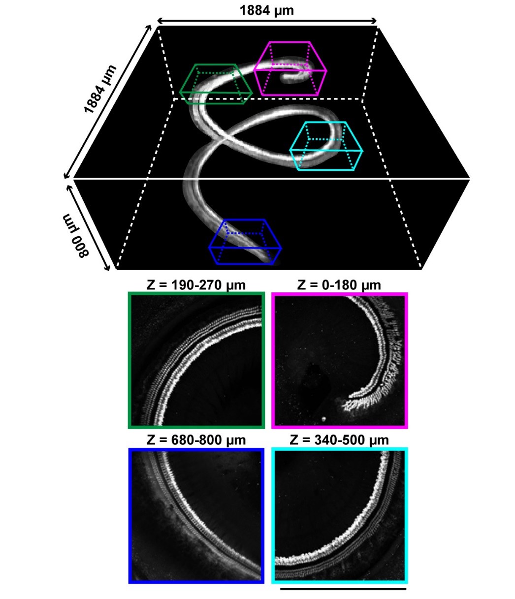
The images of the reconstructed cochlea with sensory cells identified by staining with anti-myosin 7A antibody. Cells at the depth of 800 micrometers can be detected through the bone tissue with this tissue clearing technique.
© 2019 Urata et al.
Researchers have developed a method to take high-definition images of a snail shell-shaped hearing organ located in the inner ear, called the cochlea, which converts sound vibrations into nerve impulses. The imaging technique will allow both basic and clinical scientists to study age-related and noise-induced hearing loss in more detail than ever before.
The team of researchers was led by Professor Shigeo Okabe of the University of Tokyo’s Graduate School of Medicine and included collaborators from Kyoto University's Graduate School of Medicine.
The method developed by the team — using an imaging technique called tissue clearing, which visualizes tissues and organs by making them transparent — is optimized for the cochlea, and uses machine learning-based pattern recognition to enable high-fidelity detection and analysis of all sensory cells along the entire length of the cochlea, the body's microphone.
"Sensory cells in the cochlea are small and highly packed, making the organ one of the most difficult tissue samples to be analyzed," said Okabe.
The three-dimensional structure of the organ is inevitably lost using traditional methods to dissect the tissue. The 3D structure is especially important for understanding the cochlea's function because researchers know that different areas of the organ respond to different sound frequencies and that hearing loss in human patients is frequency-dependent.
"We were motivated to attack this difficult problem by using newly developed techniques of tissue clearing and machine learning. We worked hard to optimize the software algorithm and imaging conditions, and we finally succeeded. The entire processes of image analysis and cell detection do not need any pre-tuning of the parameters," said Okabe.
After just a few days of processing, this method can visualize, record, and quantitatively analyze at single-cell resolution the millions of sensory hair cells in the cochlea.
Studying the natural 3D structure of the cochlea with this new imaging technique will reveal more details about both normal hearing and disease progression, and possibly lead to therapeutic strategies for age-related hearing loss.
Papers
Shinji Urata, Tadatsune Iida, Masamichi Yamamoto, Yu Mizushima, Chisato Fujimoto, Yu Matsumoto, Tatsuya Yamasoba, and Shigeo Okabe, "Cellular cartography of the organ of Corti based on optical tissue clearing and machine learning," eLife: January 18, 2019, doi:10.7554/eLife.40946 .
Link (Publication )
)


